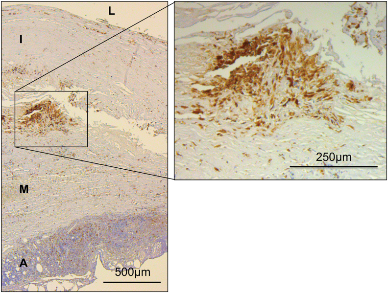
FIG. 4.
Immunohistochemical staining of macrophages in human AAA tissue. Macrophages and other CD68+ cells (i.e., SMC-derived macrophage-like cells) stained with anti-CD68 antibodies. Note CD68+ cells are accumulating mainly in the border region between tunica intima and tunica media, whereas lymphocytes are predominantly located in the adventitia forming the VALT. A, adventitia; I, tunica intima; L, vessel lumen; M, tunica media; SMC, smooth muscle cell; VALT, vascular-associated lymphoid tissue. Color images are available online.

