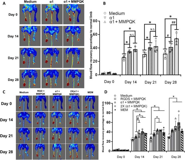
Fig. 6. Enhanced recovery of blood perfusion after ischemic hindlimb injury using α1-formulated hydrogel.
(A) Representative images of laser Doppler perfusion imaging on the ischemic hindlimb after injecting the different formulations (medium, α1 only, and α1 + MMPQK) of alginate hydrogels for serial analysis (left leg, normal; right leg, ischemic). (B) Quantification of blood flow recovery percentage of ischemic limb to normal limb (n = 4 to 5). * indicates significance difference; P < 0.05. N.S. indicates no significant difference between the groups; P > 0.05. # indicates improved blood flow of the α1 hydrogel–treated group compared to medium-treated group at day 28; P = 0.053. ## indicates the improved blood flow of α1 + MMPQK hydrogel–treated group compared to α1-treated group at day 28; P = 0.054. (C) Representative images of laser Doppler perfusion imaging on the ischemic hindlimb after injecting the formulations of the second batch (groups shown in fig. S9B) alginate hydrogels for serial analysis (left leg, normal; right leg, ischemic). (D) Quantification of blood flow recovery percentage of ischemic limb to normal limb (n = 4 to 5). * indicates significance difference; P < 0.05. # indicates the improved blood flow of α1 + MMPQK hydrogel–treated ischemic legs compared to RGDS + MMPQK hydrogel ischemic legs at day 14; P = 0.052.

