Fig. 4.
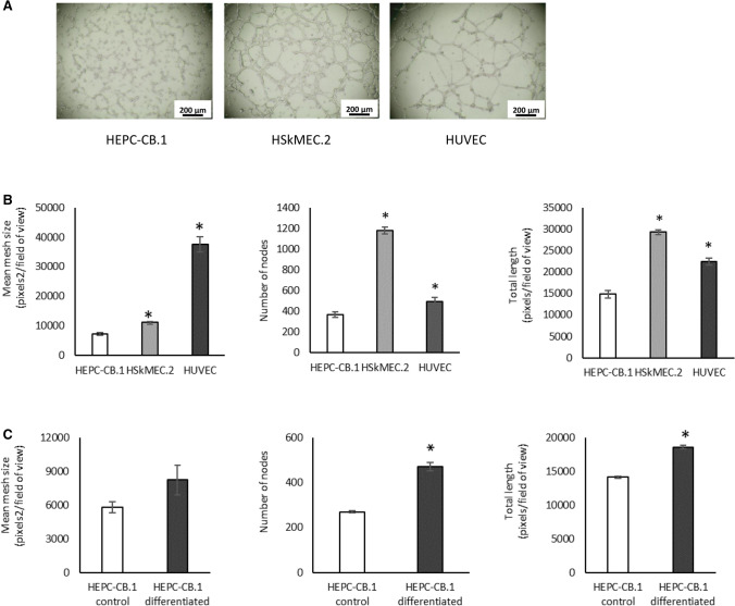
Capillary-like structure formation in Matrigel assay by HEPC-CB.1, HSkMEC.2 and HUVEC cells. A. The images show the tube formation by HEPC-CB.1 as compared to HSkMEC.2 and HUVEC cell lines, magnification ×40. B. Analysis of the results shown in A. C. Differentiation of HEPC-CB.1 cells leads to an increase in the efficiency of capillary-like structure formation. The results were obtained after 6 h of the test using the microscope Axiovert 200 M and AxioVision and ImageJ software. The values in the graphs represent the mean mesh size, number of nodes and total length ± SD of the representative experiment, determined on the basis of three independent measurements. P values were determined by t-test. *Indicates statistically significant differences (p < 0.05): in B. between HSkMEC.2 and HUVEC as compared to HEPC-CB.1 cells and in C. between differentiated and control HEPC-CB.1 cells

