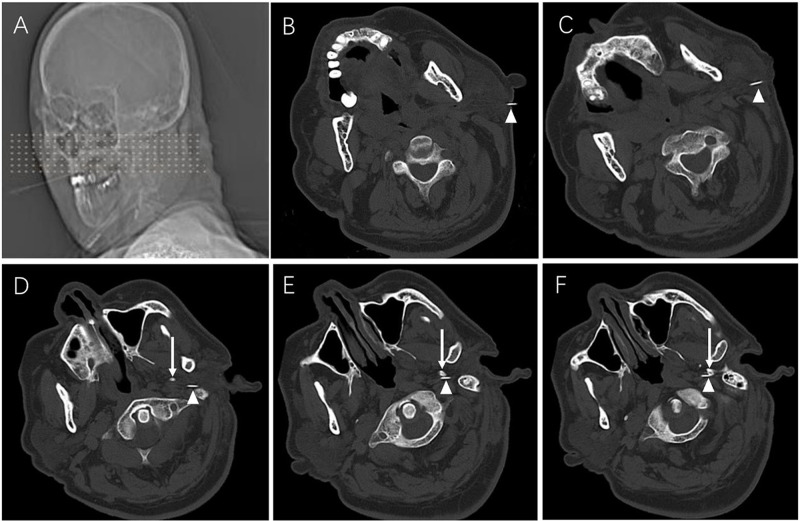
Figure 1.
PRF procedures. (A) Location for puncture. (B–F) Consecutive CT scans showing the puncture needle gradually reaching the medial edge of styloid process. Patients were placed in a supine position with head slightly turned contralaterally. White arrows indicate the needle, white triangles indicate the styloid process.

