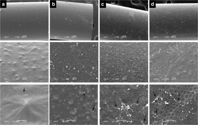
Fig. 2.
Scanning electron microscopy of polyurethane catheter samples. a Uncoated device surface (arrow indicates subtle porosity of the catheter surface). b Surface coating by the formulation film without the addition of antifungal agent (arrows indicate the covering of the catheter surface in the form of layers). c Device surface covered by formulation film containing anidulafungin (AND) 1 μg mL−1 (the arrows indicate the covering of the catheter surface in the form of very irregular networks, the pores on the surface of the device are not observed). d Catheter covered by anidulafungin/amphotericin B (AND/AMB 0.5/2.5 μg mL−1) formulation (arrows indicate the coverage of the catheter surface in the form of a very irregular mesh, the pores on the surface of the device can be observed)

