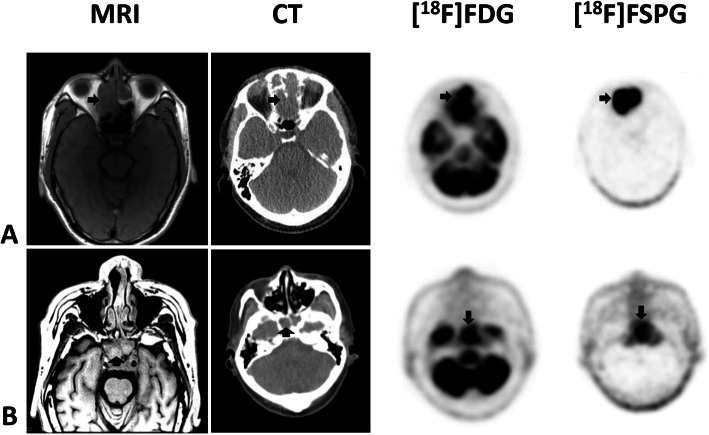
Fig. 2.
Combined image set shows two representative subjects with primary head and neck cancer (HNC). Shown from left to right are the axial T1-weighted MRI, CT, [18F]FDG PET, and [18F]FSPG PET images. The first subject (row a) has a large nasal mass while the second subject (row b) has a mass in the clivus/nasopharynx. The black arrows indicate the lesions. Any unmarked uptake on the PET scans is physiologic. All modalities show the lesions, although MRI and PET are clearer than CT. The relative uptake of [18F]FSPG and [18F]FDG within all 5 HNC subjects is similar (SUV of 2.76 ± 2.13 for [18F]FSPG and 4.92 ± 2.16 for [18F]FDG), although the complete lack of [18F]FSPG background uptake in normal brain (SUV of 0.1) versus [18F]FDG (SUV of 7.88 ± 1.45) makes evaluation of the [18F]FSPG images much easier and more suitable, for example, in radiation treatment planning

