Fig. 3.
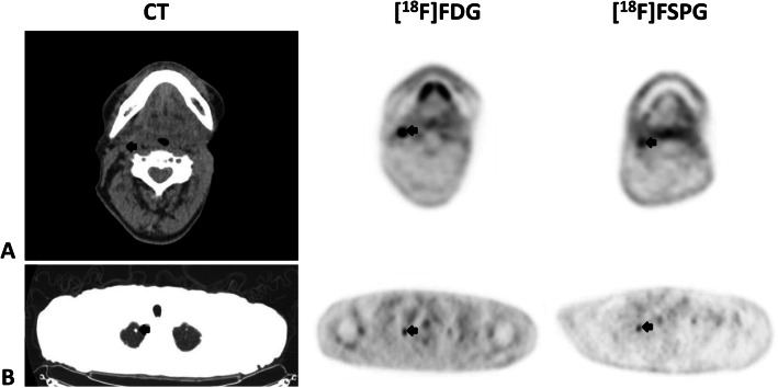
Combined image set shows two representative subjects with metastatic head and neck cancer (HNC). Shown from left to right are the axial CT (upper is soft-tissue window, lower is lung window), [18F]FDG PET, and [18F]FSPG PET images. The first subject (row a) has a right level 2 cervical lymph node, while the second subject (row b) has multiple pulmonary nodules of which a right apical nodule is shown. The black arrows indicate the lesions. Any unmarked uptake on the PET scans is physiologic. Of note, both the cervical node and the pulmonary nodule are subcentimeter in size (9 mm for the cervical node (a) and 7 mm for the right upper lobe pulmonary nodule (b)). The lesions are seen on all modalities, and both the [18F]FDG and [18F]FSPG PET images have uptake in both the cervical node (SUV of 8.7 and 2.6, respectively) and the pulmonary nodule (SUV of 2 and 1.5, respectively)

