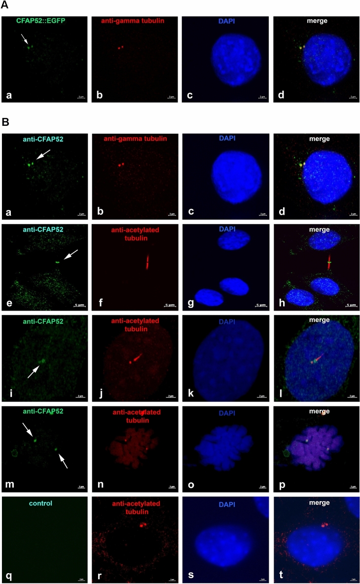
Figure 7.
CFAP52 locates to the centrosome, the basal body, the intercellular bridge, and the spindle poles in NIH3T3 cells. (A) The full-length pCfap52::Egfp expression plasmid was transfected into NIH3T3 cells and detected by auto-fluorescence (green) (a). Immuno-decoration of the centrosome with the centrosomal marker γ-tubulin (red) revealed localization of CFAP52 in the centrosome (b–d). Bars are of 2 µm. (B) Endogenous expression of CFAP52 in NIH3T3 cells was detected by double immunostaining with anti-CFAP52 (green) and either anti-γ-tubulin (red) or anti-acetylated tubulin (red). γ-tubulin and acetylated tubulin decoration for detection of the centrosome, acetylated tubulin for decoration of the basal body, the spindle poles, the intercellular bridge, and the primary cilium. The endogenous CFAP52 (arrows) locates to the centrosome (a–d), the intercellular bridge (e–h), the basal body and its associated daughter centriole (i–l), and the spindle poles (m–p). Omitting anti-CFAP52 showed decoration of the centrosome by anti-acetylated tubulin staining (red) but no false CFAP52 staining (q–t). Nuclear counterstain with DAPI (blue). Bars are of 2 µm except for (e–h), which are of 5 µm.

