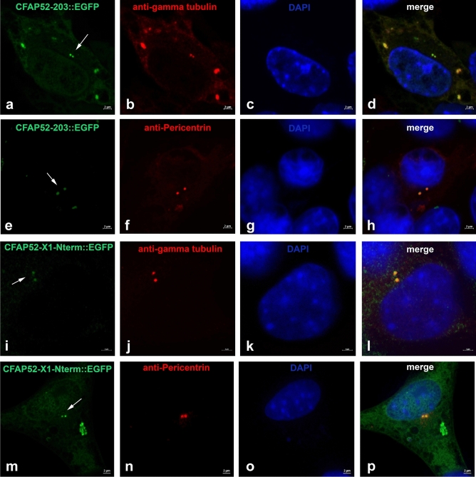
Figure 9.
Centrosomal location of EGFP-fusion proteins of CFAP52 isoforms 203 (CFAP52-203::EGFP) and the N-terminal end of isoform X1 (CFAP52-X1-Nterm::EGFP). Expression plasmids for the isoform 203, pCfap52-203::Egfp (a–h), and the N-terminal sequence of CFAP52-isoform-X1, pCfap52-X1-Nterm::Egfp (i–p) were transfected into NIH3T3 cells and detected by the EGFP auto-fluorescence (green, a, e, i, m). Decoration of the centrosomes by immunostaining for the centrosomal marker proteins either γ-tubulin (anti-gamma-tubulin, b, j) or Pericentrin (anti-Pericentrin, f, n) (both in red). Nuclear counterstain with DAPI (blue). Arrows pointing to the centrosomal location of the fusion proteins. Bars are of 2 µm (a–p).

