Fig. 1.
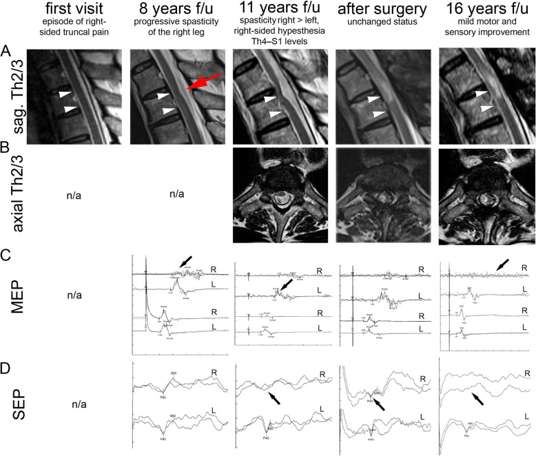
Longitudinal course of anterior spinal cord herniation. a Magnifications of the Th2/Th3 segment obtained in T2-weighted sagittal imaging at different visits. Arrowheads highlight evolving anterior spinal cord herniation and a postoperative myelopathy signal. The red arrow highlights the early subtle sign of ventral spinal cord misplacement and posterior indentation. b Corresponding axial images of Th2/3. c MEP including transcranial (lines 1–2) and lumbar stimulation (lines 3–4) to the right (R) and to the left (L) tibialis anterior muscle. Central motor conductance was pathological after 8 years follow-up (arrow; amplitude R 1.27 mV, L 2.88 mV). After 11 years, spasticity had progressed to the left leg, along with affection of the left leg central motor conductance (arrow; R 1.0 mV, L 2.47 mV). Six months after neurosurgery, there were no significant changes in MEP (R 0.68 mV, L 3.02 mV). Five years after neurosurgery, cortical MEP were absent to the right leg (arrow; R –, L 2.59 mV), despite unchanged MRI. d SEP-recordings during follow-up visits, obtained from stimulation of the right (R) and left (L) tibial nerve. At 8 years follow-up, SEP P40-latency was prolonged bilaterally (R 51.8 ms, L 52.0 ms). At presentation with Brown-Séquard syndrome involving right-sided hypesthesia, SEP from the right was absent (arrow; R –, L 50.8 ms). Six months after neurosurgery, SEP from the right gradually improved (arrow; R 51.2 ms, L 51.2 ms). Five years after neurosurgery, SEP from the right leg was absent again (arrow; R –, L 53.6 ms), despite unchanged MRI. Scale: C, 20 ms/ Div. and 2000 μV/ Div.; D, 20 ms/ Div. and 1.0 μV/ Div. f/u follow-up, n/a not available

