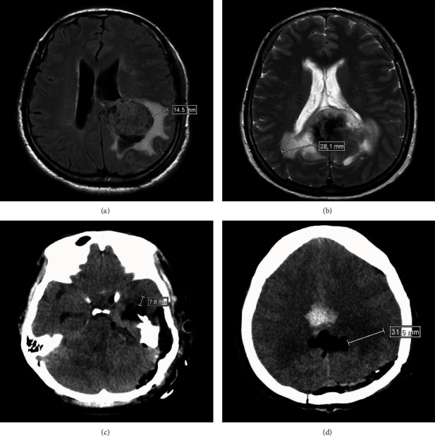
Figure 1.

Evaluation of peritumoral brain edema (PTBE) and pericavity brain edema by magnetic resonance imaging (MRI) and computed tomography (CT), respectively. (a) MRI scan showing mild PTBE before surgery. (b) MRI scan showing severe PTBE before surgery. (c) CT scan showing mild brain edema around the cavity after surgery. (d) CT scan showing severe brain edema around the cavity after surgery.
