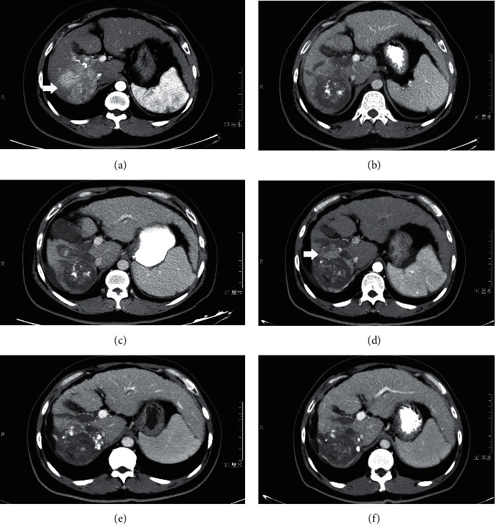
Figure 4.

Images of diagnosis and follow-up of a 37-year-old patient with massive HCC and PVTT. (a) CT showing tumor and thromboses in the right branch of portal vein (arrow). (b, c) CT showing no tumor and PVTT enhancement at 1 month and 3 months after first TACE + RFA. (d) CT showing tumor enhancement at 5 months after first TACE + RFA (arrow). (e, f) CT showing no tumor enhancement at 2 months and 5 months after second TACE + RFA.
