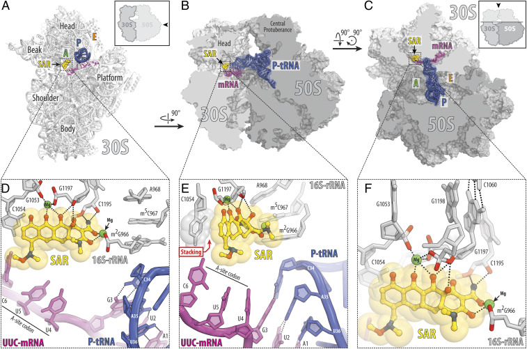
Fig. 2.
Structure of SAR in complex with the 70S ribosome, UUC-mRNA, and P-site tRNA at 2.8-Å resolution. (A–C) Overview of the SAR binding site (yellow) on the T. thermophilus 70S ribosome viewed from three different perspectives. The 30S subunit is shown in light gray, the 50S subunit is dark gray, the mRNA is magenta, and the P-site tRNA is colored dark blue. In A, the 30S subunit is viewed from the intersubunit interface, as indicated by Inset (the 50S subunit and parts of the P-site tRNA are removed for clarity). The view in B is a transverse section of the 70S ribosome. The view in C is from the top after removing the head of the 30S subunit and protuberances of the 50S subunit, as indicated by Inset. (D–F) Close-up views of the SAR interactions with the decoding center on the 30S ribosomal subunit. The E. coli numbering of the nucleotides in the 16S rRNA is used. Potential H bond interactions are indicated with dashed lines. Nucleotides of the mRNA are numbered relative to the first adenine in the P-site codon. Note that the C7 extension of SAR appears in close proximity to the third nucleotide of the A-site codon.

