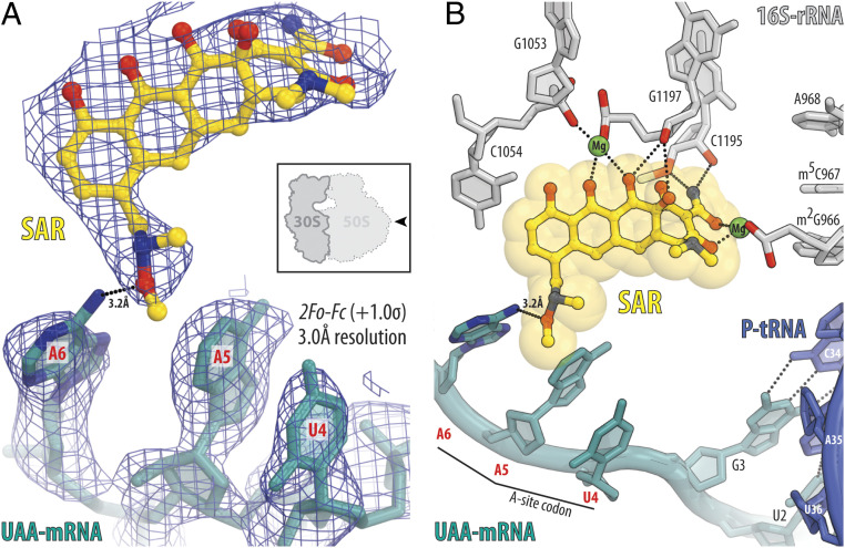
Fig. 4.
Structure of SAR in complex with the 70S ribosome, UAA-mRNA, and P-site tRNA at 3.0-Å resolution. (A) 2Fo-Fc electron density map (blue mesh) of SAR (yellow) in complex with the T. thermophilus 70S ribosome programmed with UAA-mRNA (teal). The refined model of SAR and mRNA with UAA codon in the A site are displayed in the electron density map contoured at 1.0 σ. (B) Close-up view of the SAR interactions with the mRNA and the nucleotides of the decoding center of the 30S ribosomal subunit. The E. coli numbering of the nucleotides in the 16S rRNA is used. Note that the exocyclic amino group (N6 atom) of the third adenine residue in the codon is within H bond distance (3.2 Å) from the oxygen of the C7 moiety of SAR (dotted lines).

