Fig. 2.
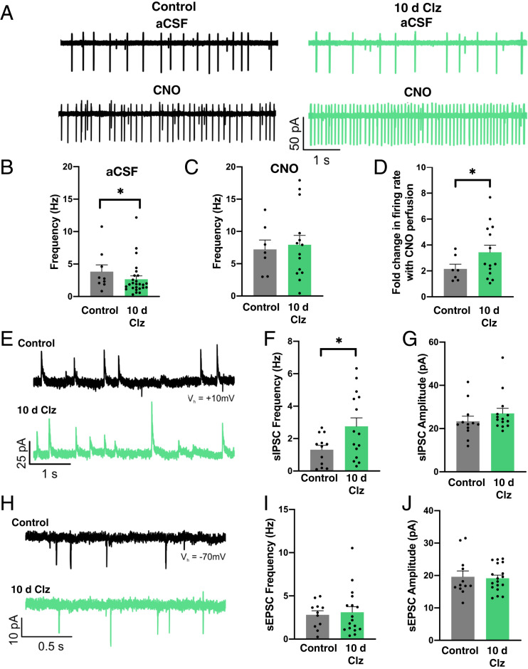
AgRP neurons remain sensitive to hM3Dq receptor activation and receive altered presynaptic input following chronic chemogenetic stimulation. (A) Representative traces showing activity of hM3Dq-expressing AgRP neurons in artificial cerebrospinal fluid (aCSF) without any CNO (upper traces) or with 5 μM CNO (lower traces) in control (Left) and Clz-treated (Right) mice. (B) Firing rate of AgRP neurons in aCSF (control: 11 cells from six mice; Clz: 26 cells from five mice; Mann−Whitney Rank Sum Test: U = 80.5, P = 0.039; *P < 0.05). (C) Firing rate of AgRP neurons in 5 μM CNO (control: seven cells from six mice; Clz: 14 cells from five mice; Student’s t test: t(19) = 0.305, P = 0.764). (D) Fold change from baseline in CNO-evoked firing rate (control: nine cells from six mice; Clz: 14 cells from five mice; Student’s t test: t(21) = 2.289, P = 0.032). (E) Representative sIPSC traces. (F) The sIPSC frequency (control: 12 cells from six mice; Clz: 15 cells from five mice; Welch’s t test: t(20.631) = −2.441, P = 0.024; *P < 0.05). (G) The sIPSC amplitude (control: 12 cells from six mice; Clz: 14 cells from five mice; Mann−Whitney Rank Sum Test: U = 70, P = 0.487). (H) Representative sEPSC traces. (I) The sEPSC frequency (control: 11 cells from six mice; Clz: 17 cells from five mice; Student’s t test: t(26) = 0.302). (J) The sEPSC amplitude (control: 12 cells from six mice; Clz: 18 cells from five mice; Student’s t test: t(28) = −0.196, P = 0.846).

