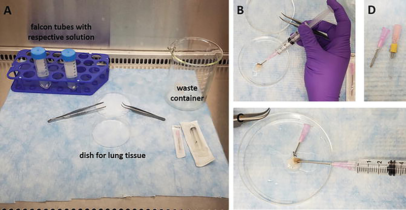
Fig. 1.
Rodent lung decellularization. (a) Hood setup with indication of the dish for the rodent lung tissue, waste container, and conical polypropylene tubes with solutions used during the decellularization; (b) flushing of a mouse lung via the tracheal cannula; (c) flushing of a mouse lung vasculature via the right ventricle; (d) cannulas used for rodent lung decellularization; left, cannula for mouse lungs, 18 gauge cannula with blunted edge and groove for attachment of the surgical thread; right, cannula for rat lungs, a silicone tube is used over a blunted 18 gauge cannula to increase diameter of the cannula and fit rat trachea size

