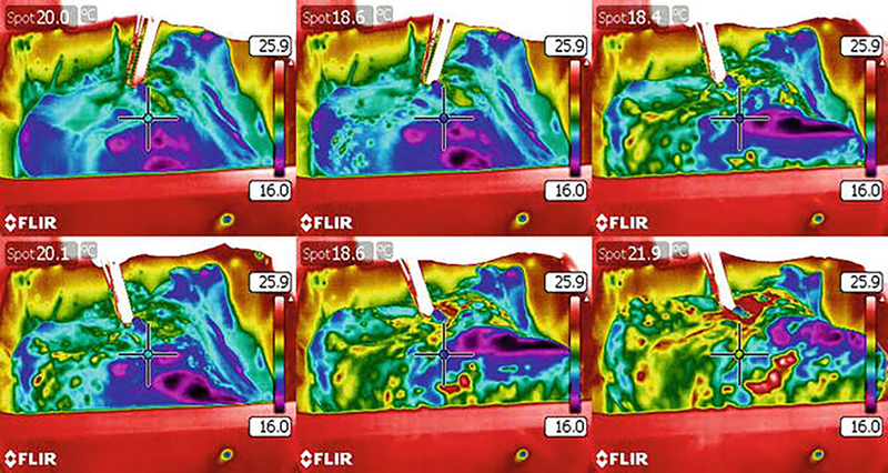
Fig. 4.
Thermographic imaging of the liquid distribution during decellularization of a human idiopathic pulmonary fibrosis (IPF) lung lobe. Representative still images from thermographic analysis demonstrate nonhomogenous liquid distribution during decellularization of a human IPF lung lobe indicated by color changes. Regions that were limited or not accessible remained blue/violet. The human lobe was chilled at 4 °C in PBS after completion of the decellularization protocol. diH2O was warmed to 37 °C to create a thermal gradient (temperature scale bar shown) for detection by the FLIR camera

