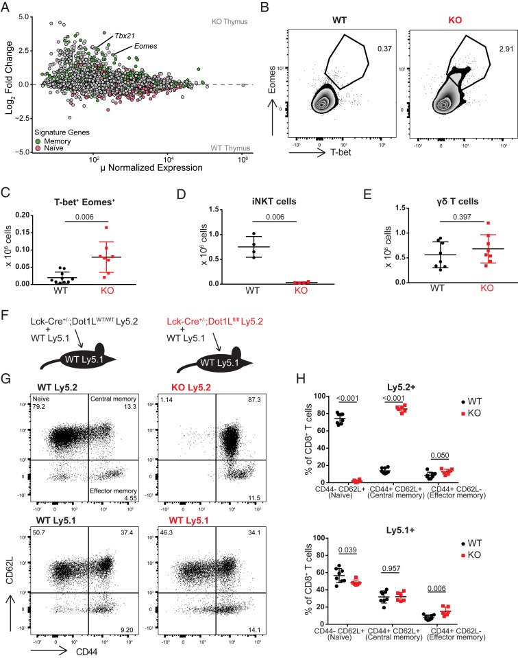
Fig. 2.
The TAIM phenotype in Dot1L-KO initiates in the thymus and is cell intrinsic. (A) MA plot of RNA-Seq data from sorted CD4−CD8+CD3+ thymocytes from three WT and four KO mice. Naïve and memory signature genes were defined as described in Fig. 1C. (B) Representative plot and (C) quantification of T-bet and Eomes expression on CD4−CD8+CD3+ thymocytes; data are of three individual experiments with two to four mice per genotype, represented as mean ± SD. (D) Quantification of iNKT (CD1d-PBS57+TCRβ+) cells in total thymus. Data are from one experiment with four mice per genotype, represented as mean ± SD. (E) Absolute number of γδTCR+ cells in spleen; data are of two individual experiments with four mice per genotype, represented as mean ± SD. (F) Outline of the mixed bone-marrow chimeras. (G and H) Representative flow-cytometry plots and quantification of CD44 and CD62L expression on Ly5.1+ and Ly5.2+ CD8+ splenocytes from mixed bone-marrow chimeras 4 mo after irradiation and reconstitution. All recipient mice were Ly5.1+ and transplanted with a mixture of either WT Ly5.1+ and Ly5.2+ Lck-Cre+/−;Dot1Lwt/wt (WT; Left) or WT Ly5.1+ and Ly5.2+ Lck-Cre+/−;Dot1Lfl/fl (KO; Right). Data are from one experiment with six or eight mice per genotype, represented as mean ± SD.

