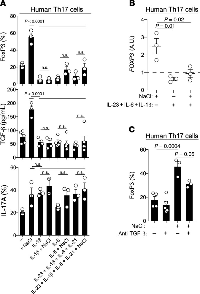
Figure 5. Proinflammatory cytokines abrogate the antiinflammatory switch in the Th17 cell phenotype that is induced in high-NaCl conditions.
(A) FACS analysis (FoxP3, IL-17A) and ELISA (TGF-β) of human Th17 cells stimulated with CD3 and CD28 mAbs for 48 hours in high- or low-NaCl conditions for a total culture period of 5 days. P < 0.0001, by 1-way ANOVA (FoxP3 and TGF-β); P = 0.03, by 1-way ANOVA (IL-17A). (B) qRT-PCR analysis of human Th17 cells on day 5 after stimulation with CD3 and CD28 mAbs for 48 hours in high- or low-NaCl conditions and the presence or absence of Th17-polarizing cytokines. P = 0.01 and P = 0.02, by 1-way ANOVA. (C) FACS analysis performed with cells treated as in B. P = 0.0004 and P = 0.05, by 1-way ANOVA. ±, in the presence or absence of; ± NaCl, in high- or low-NaCl conditions.

