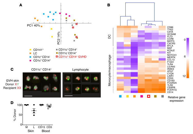
Figure 3. CD14+CD11c+ myeloid cells are donor-derived macrophages.
(A) PCA of immune gene expression by CD11c+CD14+ GVHD cells and 6 myeloid subsets from healthy control skin. Myeloid cells were sorted from healthy control skin as described in Figure 1 and are annotated accordingly. (B) Heatmap showing unsupervised clustering of CD11c+CD14+ cells from GVHD skin and myeloid cells derived from healthy control skin. Mean log2 expression for each subset is shown. n = 2 for CD141+; n = 3–6 for all other subsets. (C) Example of FISH showing the XY genotype of GVHD macrophages (CD11c+CD14+) and lymphocytes sorted from a female recipient transplanted with a male donor. A single field viewed at ×10 magnification was concatenated to show 8 representative cells per image. Scale bars: 20 μm. (D) Percentages of donor origin analyzed by XY FISH of macrophages (M) and lymphocytes (L) sorted from lesional GVHD skin compared with CD15+ myeloid cells (CD15) and lymphocytes (CD3) sorted from paired blood samples.

