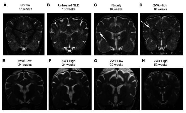
Figure 2. MRI of the brain at 1.5T.
T2-weighted images at the level of the caudate nucleus in a normal dog at 16 weeks of age (A), an untreated GLD dog at endpoint (16 weeks of age) (B), IS-only at endpoint (16 weeks of age) (C), 2Wk-High at 16 weeks of age (D), 6Wk-Low at endpoint (24 weeks of age) (E), 6Wk-High at endpoint (34 weeks of age) (F), 2Wk-Low at endpoint (29 weeks of age) (G), and 2Wk-High at 52 weeks of age (H). White arrow in C indicates widened sulcus; black asterisk in C indicates enlarged ventricle. White arrow in D indicates hyperintensity of the centrum semiovale relative to gray matter.

