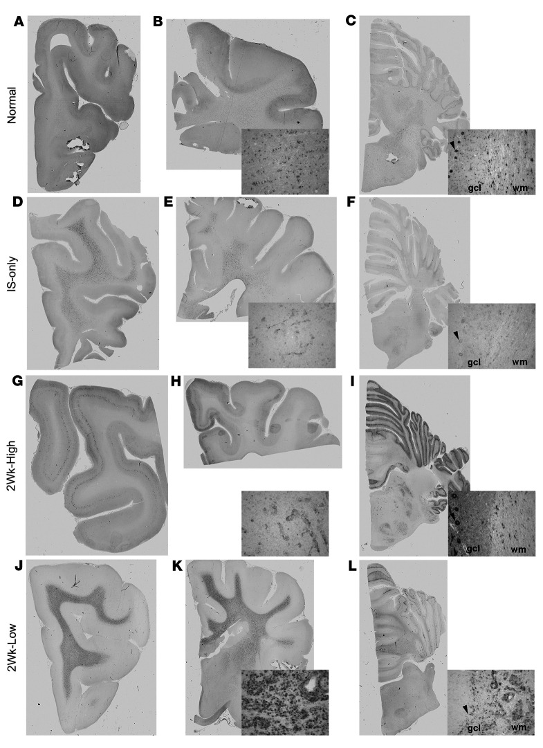
Figure 5. GALC immunohistochemical staining of the brain.
IHC staining with GALC antibody of the frontal lobe, corona radiata/internal capsule, and cerebellum in normal dog (A–C), IS-only dog (D–F), 2Wk-High dog (G–I), and 2Wk-Low dog (J–L). Insets show higher-magnification images of whole-slide scans. Black arrowheads indicate Purkinje cells. gcl, granule cell layer; wm, white matter.

