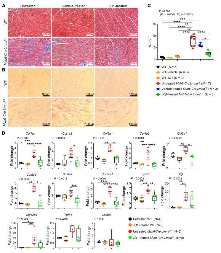
Figure 9. Effect of BET bromodomain protein inhibition on myocardial fibrosis in the Myh6-Cre LmnaF/F mice.
(A and B) Representative Masson’s trichrome–stained (top panels) and Picrosirius red–stained (bottom panels) myocardial sections after 1 week of treatment in 3-week-old untreated, vehicle, and JQ1-treated WT and Myh6-Cre LmnaF/F mice. (C) Corresponding quantitative data showing the percentage of CVF in myocardial sections in untreated (black dots; n = 5), vehicle-treated (yellow dots; n = 3), and JQ1-treated WT (orange dots; n = 3), as well as untreated (red dots; n = 7), vehicle-treated (blue dots; n = 3), and JQ1-treated (green dots; n = 6) Myh6-Cre LmnaF/F, mice. P values were calculated using 2-way ANOVA and Tukey’s post hoc test for comparisons; *P < 0.05, **P < 0.01, ***P < 0.001, ****P < 0.0001. (D) Transcript levels of selected BRD4 target genes involved in fibrosis in the heart as quantified by RT-qPCR in untreated (black dots; n = 4) and JQ1-treated (orange dots; n = 5) WT mice, as well as untreated (red dots; n = 6) and JQ1-treated (green; n = 8) Myh6-Cre LmnaF/F mice. P values shown were obtained with ordinary 1-way ANOVA or Kruskal-Wallis; *P < 0.05, **P < 0.01, ***P < 0.001, ****P < 0.0001.

