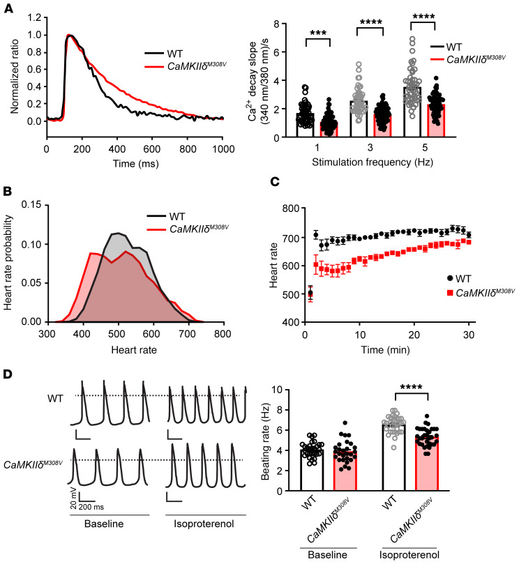
Figure 4. M308 determines fight-or-flight responses in mouse heart.
(A) Left: Representative Ca2+ transients from ventricular myocytes isolated from WT and CaMKIIδM308V mice. Right: Diastolic Ca2+ decay rates in WT and CaMKIIδM308V ventricular myocytes at various pacing frequency stimuli (WT n = 4 mice, n = 49 cells; CaMKIIδM308V n = 4 mice, n = 66–72 cells). (B) Heart rate at baseline averaged over a 24-hour period recorded by implanted telemeters in WT and CaMKIIδM308V mice (WT n = 5, CaMKIIδM308V n = 7). WT median heart rate = 520 bpm, CaMKIIδM308V median heart rate = 505 bpm (P < 0.0001). (C) Heart rate responses to isoproterenol injection (0.4 mg/kg/mouse, intraperitoneal) in WT compared with CaMKIIδM308V mice (WT n = 5, CaMKIIδM308V n = 7). (D) Left: Representative tracings of beating rate of isolated sinoatrial cells from WT and CaMKIIδM308V mice at baseline and after isoproterenol treatment (right panel). Right: Quantitative analysis of beating rates (WT n = 6 mice, n = 28–33 cells; CaMKIIδM308V n = 7 mice, n = 28–33 cells). ***P < 0.001, ****P < 0.0001 by 1-way ANOVA with Tukey’s multiple-comparisons test (A and D).

