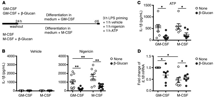
Figure 1. Effect of β-glucan on IL-1β release upon NLRP3 inflammasome activation.
(A) Schematic overview of innate immune memory methodology. Monocytes were preincubated with β-glucan or left untreated in a medium containing either GM-CSF or M-CSF. After 24 hours, the stimulus was washed away, and the cells were differentiated for an additional 5 days, after which the macrophages were restimulated. d6, day 6. (B) Macrophages were primed for 3 hours with LPS and then stimulated for 1 hour with nigericin or vehicle as a control. Culture supernatants were collected, and the concentration of secreted IL-1β was determined by ELISA. (C) Macrophages were primed for 3 hours with LPS and stimulated for 1 hour with ATP. Culture supernatants were collected, and the concentration of secreted IL-1β was determined by ELISA. Note that the highest IL-1β values in control (None) GM-CSF and M-CSF macrophages were from different healthy donors. (D) Real-time quantitative PCR (RT-qPCR) of IL1B mRNA in macrophages primed for 3 hours with LPS. Results were normalized to β-actin expression levels. The results obtained for the cells differentiated with GM-CSF in the absence of β-glucan were arbitrarily set at 1 to express the results as fold change. (B–D) Graphs show the mean ± SEM of at least 3 independent experiments. For B, n = 10; for C and D, n = 7; *P < 0.05 and **P < 0.01, by Wilcoxon matched-pairs, signed-rank test.

