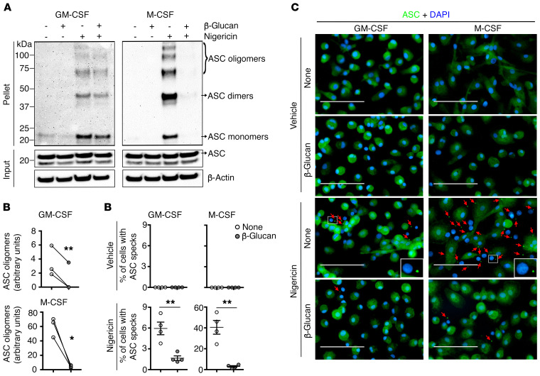
Figure 4. Effect of β-glucan on ASC oligomerization and speck formation.
(A) Monocytes were preincubated or not with β-glucan and differentiated with either GM-CSF or M-CSF. Macrophages were primed for 3 hours with LPS and then stimulated for 1 hour with nigericin or vehicle as a control. Total cell lysates were obtained in Triton X-100–containing buffer. Insoluble (pellet) fractions were cross-linked with DSS to capture ASC oligomers. The soluble (input) and insoluble fractions were analyzed by immunoblotting with an ASC Ab. β-Actin was used as a loading control. Blots are representative of 3 independent experiments. (B) Densitometric analysis of the ASC oligomers on the blots of 3 healthy donors. *P < 0.05 and **P < 0.01, by paired, 2-tailed Student’s t test. (C) Representative immunofluorescence microscopic images of ASC specks. Macrophages (generated as in A) were stimulated for 1 hour with nigericin or vehicle. DNA is stained in blue and ASC in green. Red arrowheads point to ASC specks, and the enlarged inserts show cells with an ASC speck. Original magnification, ×40. Scale bars: 100 μm. (D) Quantification of the percentage ASC specks (4 × 200 cells/nuclei [DAPI-stained], analyzed with ImageJ). Data represent the mean ± SEM of the analysis of 3 independent experiments. n = 4. **P < 0.01, by paired, 2-tailed Student’s t test.

