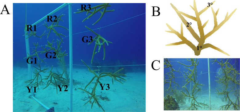
Figure 1. Nursery-reared Acropora cervicornis sampled in December 2017 for microbial community composition.
(A) Three colonies each of red (R), green (G), and yellow (Y) coral genotypes were sampled. (B) On each colony, three replicate samples were collected on basal branches (1°), intermediate branches (2°), and apical colony tips (3°). (C) The same colonies photographed in October 2018 showed rapid growth and no signs of disease. Acropora cervicornis icon courtesy of the Integration and Application Network, University of Maryland Center for Environmental Science (ian.umces.edu/symbols/).

