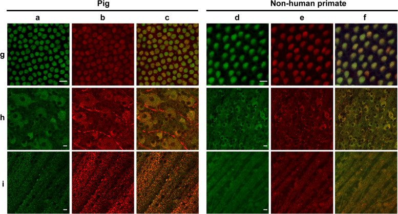
Fig. 6. Autofluorescence and mitochondrial colocalization on the flat-mounted retina from pig and nonhuman primate (Macaca fascicularis).
Autofluorescence (a, d) and mitochondrial ATP-synthase immunolabelling (b, e) with the merged images (c, f) in cone inner segments (g), ganglion cell layer (h), and optic fiber layer (i) showing co-localization except for the unspecific red labeling of blood vessels in the pig ganglion layer (h). Scale bars represent 10 µm.

