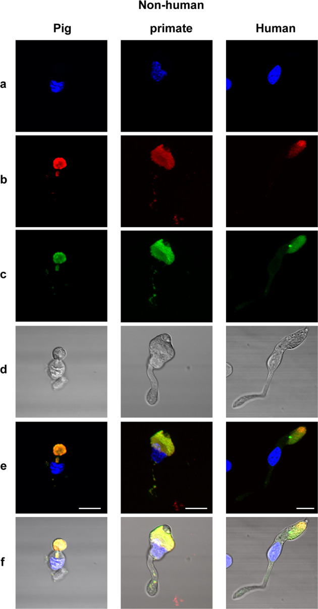
Fig. 7. Autofluorescence and mitochondria localization on isolated cone photoreceptors from pig, nonhuman primate (Macaca fascicularis), and human retina.

DAPI stained nuclei (a), mitochondrial ATP-synthase immunolabelling (b), autofluorescence (c), transmitted light in isolated cone photoreceptors, showing the autofluorescence and ATP-synthase labeling co-localization above the cell body (e, f). Note that some mitochondria are also seen in the axon of the photoreceptors on the merged images (e, f). Scale bars represent 10 µm.
