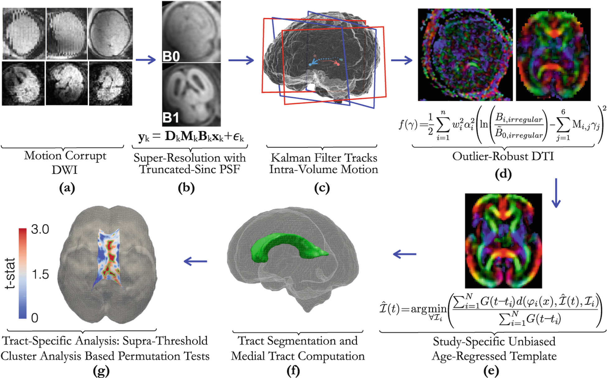
Fig. 1.

Proposed fetal brain DTI group analysis pipeline: The acquired DWI data (often multi-planar), (a) is used to compute composite super-resolved B0 and B1 images (b). Motion-tracking based slice-to-volume registration (c) is used to map b = 0 and b ≠ 0 slices to standard space [9] where DTI (d) is computed directly from motion-corrected data (shown next to a color FA image obtained directly from the scanner without motion correction). Template construction (e) provides an unbiased spatial frame where group statistical differences (f) are computed and localized (g).
