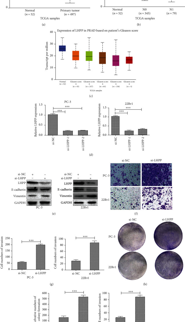
Figure 7.

Silencing of LHPP promotes the EMT, invasion, and proliferation of prostate cancer cells. (a) LHPP expression profile in PCa and normal tissues was gained from TCGA database. p < 0.01. (b) LHPP expression profile in normal tissues, N0 PCa, and N1 PCa. N0-vs-N1: p < 0.01; normal vs. N1: p < 0.01. N0: no regional lymph node metastasis; N1: metastases in 1 to 3 axillary lymph nodes. (c) LHPP expression profile in normal tissues and Gleason score PCa. Gleason score 6 vs. Gleason score 9: p < 0.01; Gleason score 7 vs. Gleason score 9: p < 0.05; Gleason score 8 vs. Gleason score 9: p < 0.05. (d) Relative LHPP mRNA expression in PC-3 and 22Rv1 cell lines transfected with si-NC, si-LHPP-1, and si-LHPP-2. Si-LHPP-1 was used in follow-up experiments. (b) Relative LHPP, E-cadherin, and Vimentin protein expression in cell lines transfected with si-NC or si-LHPP was determined by Western blotting. (c) The invasion of 22Rv1 and PC-3 cell lines transfected with si-NC or si-LHPP was determined by transwell assays. (d) Relative cell numbers of invasion were calculated. (e) The colonizing ability of 22Rv1 and PC-3 transfected with si-NC or si-LHPP was determined by colony formation assays. ∗p < 0.05, ∗∗p < 0.01, and ∗∗∗p < 0.001.
