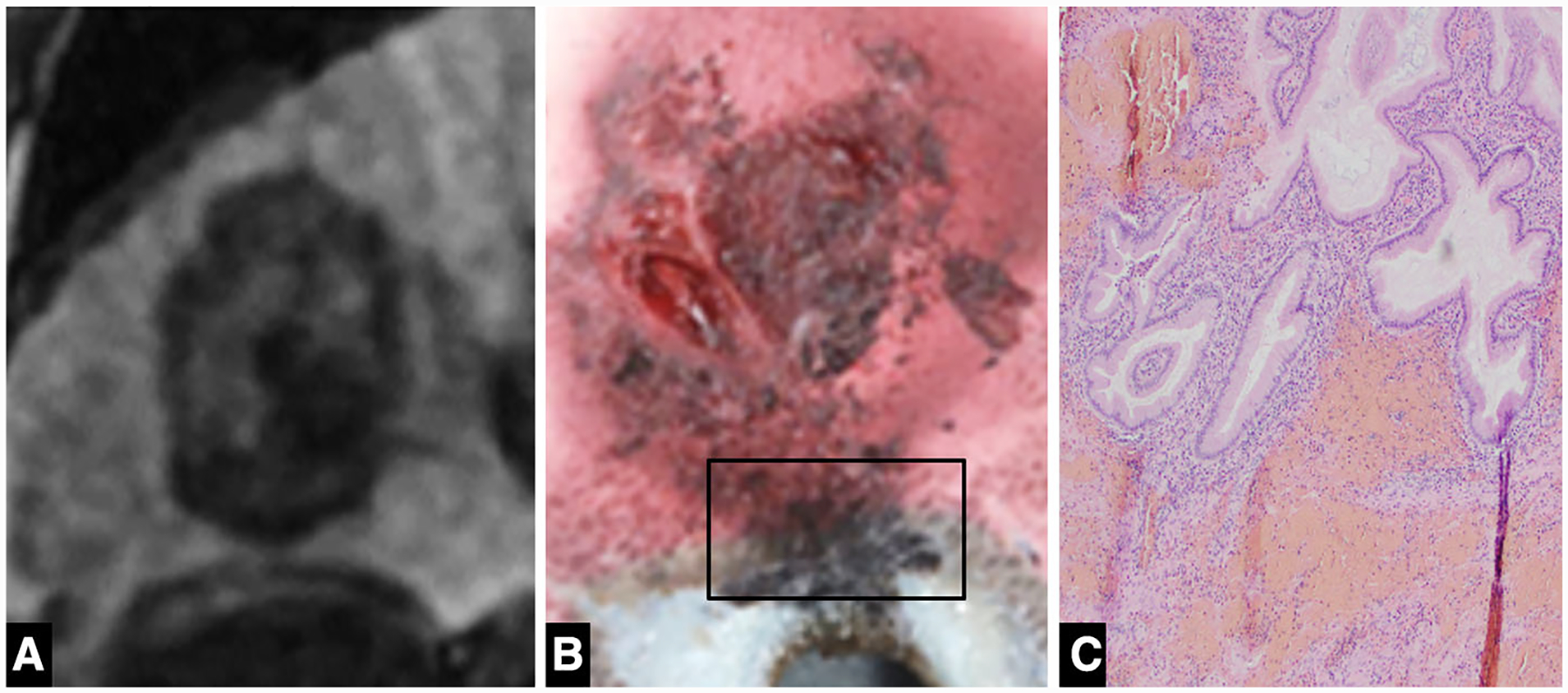
Fig. 2. A.

A Coronal 20-min delay MRI image demonstrating the close proximity of the ablation zone to the cranial aspect of the gallbladder. B Gross pathologic specimen demonstrates extension of the ablation zone to involve the gallbladder fossa. C Histology (× 100) demonstrates hemorrhage within the gallbladder wall with intact mucosa
