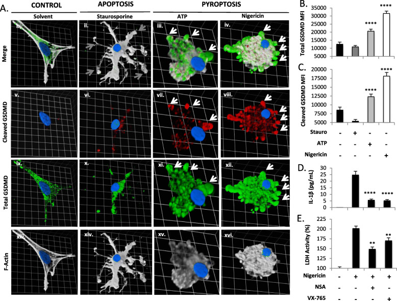
Fig. 2.
Cleaved GSDMD is present in human microglia during pyroptosis. Primary human microglia were exposed to the pro-pyroptotic stimulus ATP (100.0 μM, 24 h), the pro-pyroptotic stimulus nigericin (5.0 μM, 4 h), the pro-apoptotic stimulus staurosporine (5.0 μM, 4 h), or vehicle [equivalent volume PBS (24 h)]. Cells were fixed, immunolabelled for cleaved GSDMD (Av–viii, red) and total GSDMD (Aix–xii, green), labeled with F-actin (Axiii–Axvi, white; merge shown in Ai–iv), and visualized by confocal microscopy. Images represent three-dimensional z-stacks incorporating 15 XY planes over a vertical distance of 4–6 μm. One square unit represents 10.28 μm. (b, c) Mean fluorescence intensity (MFI) of each protein was assessed for a minimum of n = 20 microglia per condition. Cell supernatants from microglia exposed to nigericin, ATP, or staurosporine as in (a) were used to measure IL-1β release (d) and LDH activity (e) in each condition. Data shown represent mean ± SEM for a representative human donor. Data were tested for significance using one-way ANOVA with Dunnett’s test for multiple comparisons (**p < 0.01, ****p < 0.0001). All experiments were recapitulated in microglia derived from two to three separate human donors

