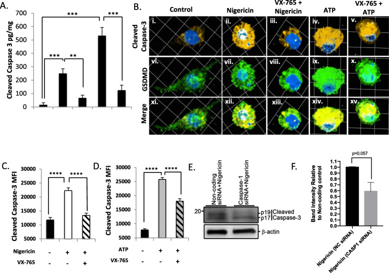
Fig. 7.
Caspase-3 activation occurs downstream of canonical caspase-1 inflammasome activation. (a) Microglia were pre-treated with VX-765 (50 μM, 4 h) and then exposed to pyroptotic stimuli ATP (100.0 μM, 24 h) or nigericin (5.0 μM, 4 h), from which lysates were harvested and cleaved caspase-3 levels assessed by ELISA. Data shown represent mean cleaved caspase-3 levels ± SEM (n = 6 technical replicates) from a representative human donor. Results were confirmed in microglia from 3 to 5 donors. (b) Microglia were exposed as in (a) and immunolabelled for cleaved caspase-3 p17/p19 (amber) and GSDMD (green). Images represent three-dimensional z-stacks incorporating 15 XY planes over a vertical distance of 4–6 μm. (c, d) Mean fluorescence intensity (MFI) of cleaved caspase-3 was assessed in control microglia (minimum n = 20) and nigericin- or ATP-exposed microglia ± VX-765 (minimum n = 100). Data shown represent mean cleaved caspase-3 MFI ± SEM for a representative human donor. Results were independently verified in microglia from 2 to 3 separate donors. (e, f) Microglia were transfected with either universal non-coding siRNA (NC siRNA) or a cocktail of three different siRNAs targeting caspase-1, exposed to nigericin, and cell lysates immunoblotted for caspase-3 as indicated. Data shown are mean protein band intensities ± SEM (n = 2 biological replicates; one-tailed Student’s t test) normalized to beta-actin, expressed relative to the untreated NC control. P17 and p19 bands were quantified collectively to quantify the total reduction in cleaved caspase-3 peptide

