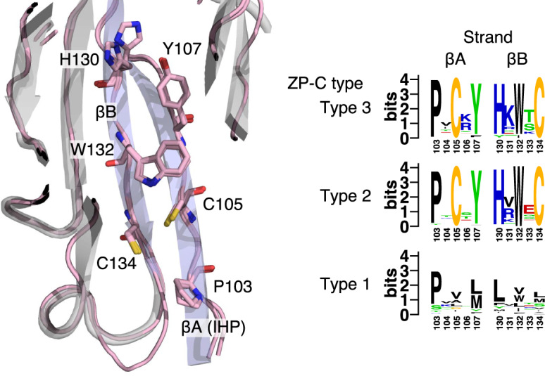
Fig. 4.

The conserved IHP of Type 2/3 ZP-C domains. Homology models for the ZP-C domains of Caenorhabditis elegans CUT-1 (Type 3) and DPY-1a (Type 2) (both shown in pink lines, with key residues shown in stick format) were superimposed on the cartoon structure of the template, human uromodulin (gray cartoon, with the βA and βB strands colored blue). The residues along the inward facing side of βA comprise the IHP; those residues, and the three adjacent residues in βB, are highly conserved in nematode Type 2 and 3 ZP-C domains and suggest a novel disulfide bond. These same sites are variable in Type 1 ZP-C domains. Conservation patterns for the three ZP-C domain types are shown via sequence logos (extracted from supplementary fig. 4, Supplementary Material online).
