
Figure 4: ERMS and ARMS can be distinguished based on histopathology features.
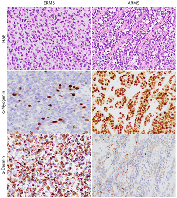
Representative photomicrographs of embryonal RMS (ERMS; left panels) and alveolar RMS (ARMS; right panels) following staining with hematoxylin and eosin (H&E; top panels) or with primary antibodies to detect Myogenin (middle panels) or Desmin (bottom panels) to mark the skeletal muscle lineage. ARMS often, but not always, displays loosely associated tumor cells in clusters resembling pulmonary alveoli and robust immunohistochemical staining for Myogenin; however, confirmation by analysis of PAX3–FOXO1 or PAX7–FOXO1 fusion is required to confirm FP state. Original magnification: 400x (Image provided by D. Rakheja, University of Texas Southwestern Medical Center)(contact information: dinesh.rakheja@utsouthwestern.edu)