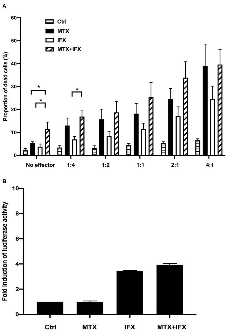
FIGURE 3.
Antibody-dependent cell-mediated cytotoxicity and antibody-dependent cellular phagocytosis induced by IFX in co-stimulation with MTX. (A) TmTNF-expressing Jurkat cells were used as target cells and labeled with membrane dye PKH26. Peripheral blood mononuclear cells (PBMCs) were used as effector cells and co-incubated with target cells on flat-bottom plates. The ratios of effector cells and target cells was from 1:4 to 4:1. Cells were incubated with MTX for 24 h, IFX for 2 h, or combination of MTX and IFX for 2 h after MTX for 24 h. As a control, target cells were incubated with effector cells without MTX and IFX. Treated cells were stained with TO-PRO-3 iodide. TO-PRO-3 iodide-positive dead cells in PKH26-positive target cells were detected by flow cytometry. Proportion of dead cells in target cells was indicated. Values are mean ± SEM; n = 4/group. (B) TmTNF Jurkat cells (2 × 10⁴ in 150 μl/well) were co-incubated with FcγRIIa-H effector cells (2 × 10⁴ in 150 μl/well) and stimulated with 0.1 μM MTX, 0.01 μM IFX, or MTX+IFX for 6 h. Treated cells were cultured with luciferase assay buffer for 20 min and the luminous cells were detected by a luminometer. Values are mean ± SEM; n = 3/group. *p < 0.05, Mann-Whitney U tests.

