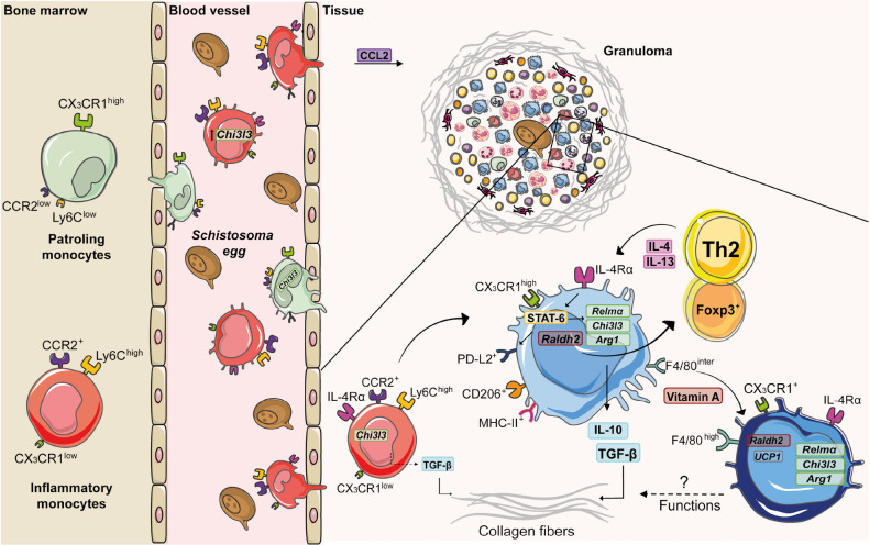
FIGURE 2.
Monocyte and macrophage dynamics during experimental S. mansoni infection. Along the course of experimental S. mansoni infection in mice, bone marrow increases the monopoiesis and originates two subsets: inflammatory monocytes (Ly6ChighCCR2highCX3CR1low) and patrolling monocytes (Ly6ClowCCR2lowCX3CR1high), which are recruited from blood to tissues affected by egg accumulation. Upon activation by circulating eggs, inflammatory monocytes express higher levels of chitinase-like 3 (Chi3l3) compared to patrolling monocytes. CCL2 mediates inflammatory monocyte recruitment to the liver during schistosomiasis. Inside the tissue, inflammatory monocytes produce TGF-β, induce collagen deposition and differentiate into macrophages. The diversity of cells present in the granuloma is responsible for a type 2 microenvironment, whereby T cells produce IL-4 and IL-13 that induces the alternative activation of macrophages via IL-4Rα. The IL-4/IL-13/IL-4Rα axis leads to the transcription of retinaldehyde dehydrogenase 2 (Raldh2) and activation of signal transducer and activator of transcription 6 (STAT-6). This transcription factor upregulates the expression of Chi3l3, programmed cell death ligand 2 (Pdl2), arginase -1 (Arg1) and resistin-like alpha (Relma), which trigger TGF-β production and collagen deposition. RALDH2 by AAM induces Treg cell differentiation. Vitamin A mediates conversion of monocyte-derived macrophages (F4/80intCD206+PD-L2+MHC-II+) into F4/80highCD206– PD-L2– MHC-II– UCP1+ phenotype, whose function still needs to be elucidated.

