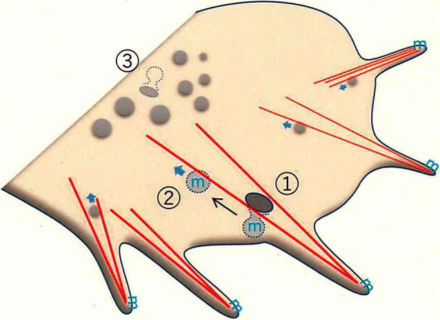
Figure 3.

Membrane trafficking in the growth cone, as revealed by super-resolution microscopy. F-actin-dependent endocytosis occurs in the peripheral (P-) domain of the growth cone. The leading edge protrudes as filopodia, which have dense F-actin bundles (red lines). F-actin-bundling for filopodial formation induces endorphin-mediated endocytosis (EME; ①; Nozumi et al., 2017). EME depends on F-actin located in the Z-axis direction (see Igarashi et al., 2018). GPM6a (the symbol “m” in blue), distributed in the lipid rafts, is endocytosed through EME. The EME-dependent vesicles move in a retrograde direction to the central (C-) domain of the growth cone (②). Classical clathrin-mediated endocytosis (CME) mainly occurs at the bottom of the growth cone membrane (GCM; ③).
