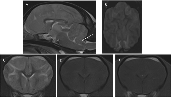
Figure 1.
Images of the first MRI study performed at presentation. (A) Mid-sagittal T2W, (B) dorsal T2W, (C) transverse, T2W, (D) T1W pre-contrast, (E) transverse T1W post-contrast. The transverse images are at the level of the nucleus caudatus, and the dorsal image is a slice taken just dorsal to the corpus callosum. The arrow in (A) points to the caudo-ventral aspect of the cerebellum, which is displaced caudally and is partially herniating through the foramen magnum. In (B), notice the blurring of the cerebral sulci, indicating increased brain volume, and the diffuse increased signal intensity of the white matter, which is also seen in (C). The signal intensity changes of white matter are barely perceptible in T1W (D,E) shows a lack of contrast enhancement.

