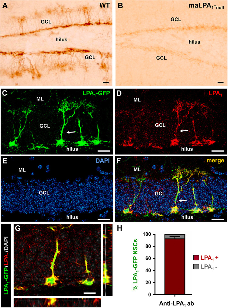
FIGURE 1.
Lysophosphatidic acid receptor 1–GFP expression colocalizes with LPA1 immunostaining and specifically labels hippocampal NSCs. Immunostaining with an anti-LPA1 antibody labels NSCs in the DG (A). When the same antibody is used in the maLPA1-null mouse, in which only a truncated non-functional form of LPA1 is expressed, staining is almost absent (B). Immunostaining with the anti-LPA1 antibody in slices from the LPA1-GFP transgenic mouse shows almost total colocalization. Confocal microscopy imaging was used to analyze colocalization in z-stack projections of the LPA1-GFP signal (C) with the anti-LPA1 immunostaining (D). DAPI staining was used for better anatomical resolution (E). The merged image is shown in (F), and an orthogonal projection demonstrating full colocalization is shown in (G). The quantification showing the almost 100% colocalization of LPA1-GFP and LPA1 immunostaining in NSCs is shown in (H). Scale bar is 20 μm in all images.

