Figure 1.
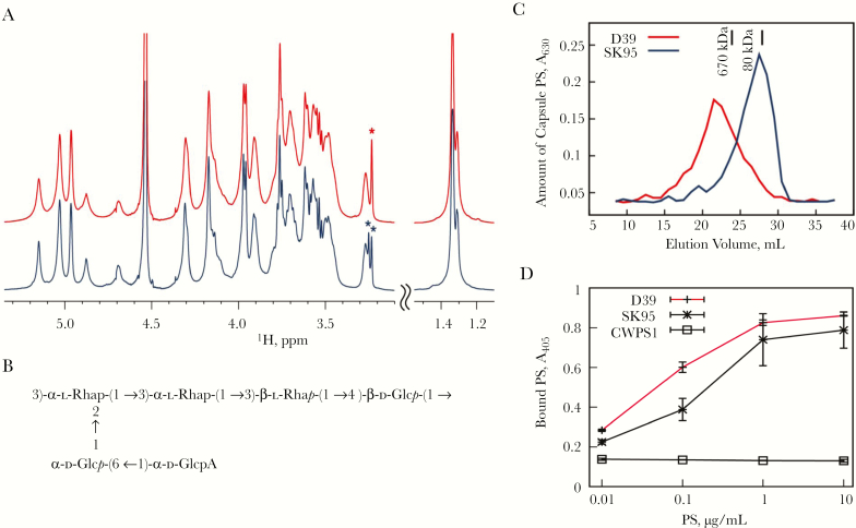
Structure of type 2 capsule polysaccharide (PS). A, Overlay of partial (1H) Nuclear magnetic resonance (NMR) spectra of capsular PSs from type 2 Streptococcus pneumoniae (D39) (red, top) and Streptococcus oralis (SK95) (blue, bottom) recorded at 50°C. As shown, spectra are identical, but the signals of D39 are broader than those of SK95. Asterisks mark choline signals arising from teichoic acid. To facilitate presentation, the 2 spectral regions are plotted with different vertical scales. B, Structure of capsular PS from S. oralis (SK95) as determined with NMR spectroscopy. C, Amount of capsular PSs from SK95 (blue) and D39 (red) versus elution volumes of an S-500 column. Elution volumes of dextran molecular weight standards are indicated with vertical bars. D, Amount of type 2 capsule (y-axis) bound to the enzyme-linked immunosorbent assay (ELISA) plate at different amounts of PS (x-axis). ELISA wells were coated with antiphosphocholine antibody and captured PSs were determined with a monoclonal antibody (mAb) specific for type 2 PS (Hyp2M2). The mAb supernatant was diluted at 1:320, 1:160, and 1:80, respectively, for D39, SK95, and CWPS1 (teichoic acid) wells. Error bars at the data points indicate standard deviations (error bars for CWPS1 were too small to be seen). Abbreviations: A405, absorbance at 405 nm; A630, absorbance at 630 nm.

