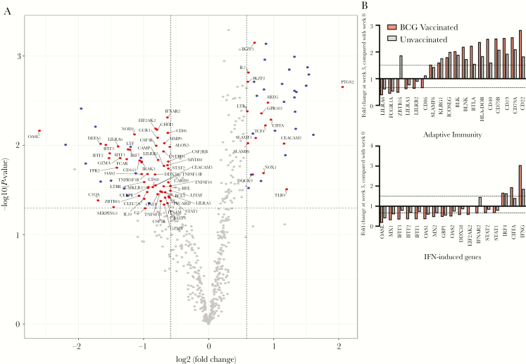
Figure 4.
Transcriptional changes in immune genes associated with BCG vaccination. Differential expression of 770 immune genes in infant macaque peripheral blood mononuclear cells (PBMCs) was assessed 0 weeks and 3 weeks after BCG vaccination. A, Changes from baseline (ie, week 0) to week 3 after BCG vaccination are shown in volcano plots for BCG-vaccinated infants (n = 6). Colored points represent genes whose expression significantly increased or decreased by >1.5-fold (P < .05; dashed lines), with blue points indicating genes with levels that were reduced or elevated in all infants and red points indicating genes that were only changed in BCG-vaccinated infants. B and C, Genes labeled in panel A with increased or decreased expression within adaptive immunity (B) and interferon-related (C) signaling pathways are shown with fold changes for BCG-vaccinated (red bars) or unvaccinated (gray) bars. Statistical measurement of differentially expressed genes is described in the Methods. IFN, interferon.

