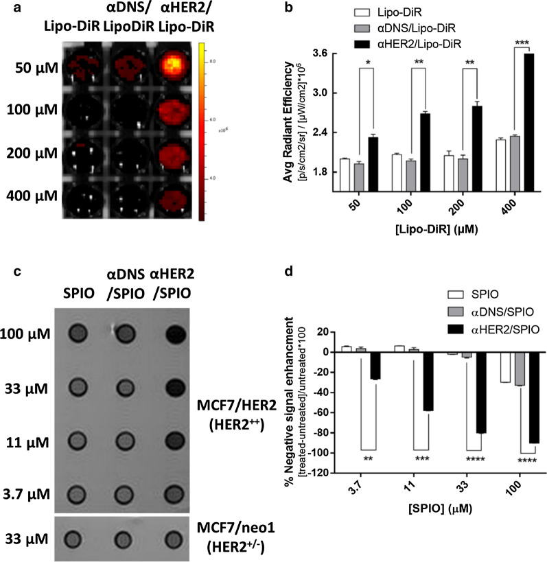
Fig. 4.
In vitro sensitivity image of αHER2/PEG-NPs. MCF7/HER2 (HER2++) cancer cells incubated with HER2 targeted-contrast agent with serial dilution concentrations. a αHER2/Lipo-DiR, αDNS/Lipo-DiR and Lipo-DiR were added to cells. Fluorescence images were obtained by the IVIS spectrum system. b Calculations of average radiant efficiency of (a). c αHER2/SPIO, αDNS/SPIO and SPIO were added to cells. MR imaging was performed with a 7.0 T MR imaging scanner. d The result from (c) was calculated by [treated SI-untreated SI]/untreated SI*100. *P < 0.05, **P < 0.01, ***P < 0.001, ****P < 0.0001 (unpaired t test)

