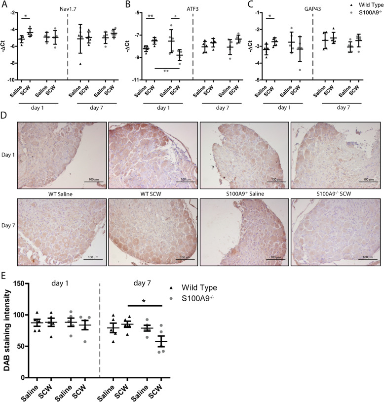
Fig. 6.
Of a panel of activation markers, ATF3, NAV1.7, and GAP43 are differentially expressed in DRG of arthritis WT versus S100A9−/− mice. Three neuronal activation markers showed a significant increase in WT mice after SCW injection: NAV1.7 (a), ATF3 (b), and GAP43(c). These differences were found only 1 day after the injection of SCW. In S100a9−/− mice, no increase in these markers were found, for ATF3 even a small decrease. At day 7, protein level NAV1.7 also seemed lower in S100a9−/− compared to saline-injected mice, whereas in WT mice, the expression seemed increased (d; magnification × 200). This was quantified, and protein expression was lower at day 7 in S100a9−/− mice, compared to WT (e). Significance was tested using a two-way ANOVA at n = 5 per group. A Tukey post hoc test was performed to identify significantly different means (*p < 0.05; **p < 0.01; ***p < 0.001)

