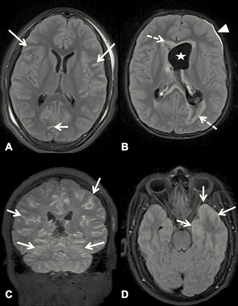
Figure 1.
Postcontrast T2-weighted fluid-attenuated inversion recovery magnetic resonance images of patients 5, 14, 19, and 8 (A–D, respectively). (A) Axial image showing abnormal leptomeningeal hyperintensity (arrows) throughout both cerebral hemispheres, consistent with meningitis. (B) Axial image showing dilated left ventricle (star), abnormal signal in periventricular white matter (dotted arrows), and left subdural enhancement (arrowhead), sequelae caused by meningitis. (C) Coronal image showing diffuse abnormal hyperintensity in the leptomeninges of the cerebellar and cerebral hemispheres (arrows). (D) Axial image showing abnormal hyperintensity (arrows) of the left temporal lobe meninges and cortex, consistent with meningoencephalitis.

