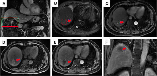
Figure 2.
Contrast-enhanced magnetic resonance imaging (MRI) revealed a 54-years-old man with HCC of 3.7 cm in maximum diameter in segment 7 who underwent microwave ablation (MWA). (A) MRI axial scan showed a residual liver in portal phase after 1 month underwent hepatectomy, the red frame shows the cut into parts; (B) a slightly higher signal nodule in red circle was shown in segment 7 in MRI T2WI (red arrow), which was defined as a recurrence lesion; (C) a high signal nodule is shown in arterial phase axial MRI image after MWA (red arrow); (D) high-density MWA zone is shown in T1WI axial MRI image after 3 months (red arrow); (E) low-density MWA zone is shown in delay phase axial MRI image after 3 months (red arrow); (F) low-density MWA zone is shown in delay phase coronal MRI image after 3 months (red arrow).

