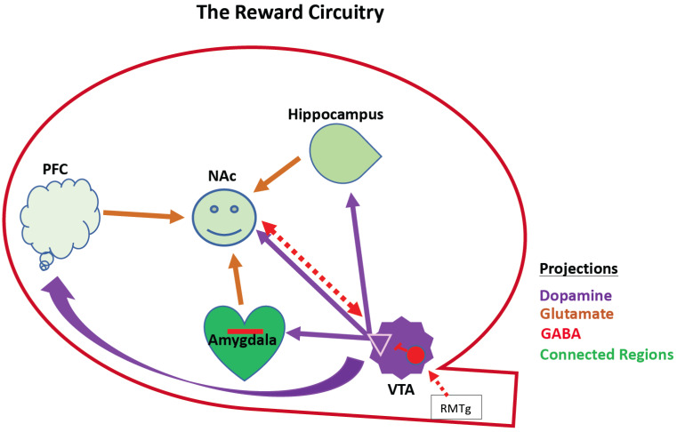
Fig. (1).
Schematic of reward circuitry. This shows interconnected mesocorticolimbic regions that interact to control reward behavior. These include the hippocampus, the amygdala, the VTA, the PFC, and the NAc. This is a VTA-NAc centric cartoon, but there are reciprocal interactions between the PFC, Hippocampus, and amygdala (not shown). Except for the RMTg, downstream components of the Brain Reward Cascade are omitted for simplicity. Dopaminergic neurons emanate from the VTA (purple). The NAc receives its dopaminergic input from the VTA and integrates that with cortical hippocampal and amygdaloid afferents to effect dopamine reward (converging arrows in NAc). NAc-VTA share reciprocal inhibitory projections and the VTA receives inhibitory afferents from the RMTg that modulate its activity. Opioid receptors modulate GABA neuron activity (red). PFC, prefrontal cortex; NAc, nucleus accumbens; VTA, ventral tegmental area; Red interneuron in the VTA represents GABAergic neurons/synapses expressing opioid receptors. NAc, Nucleus Accumbens; PFC, prefrontal cortex; VTA, ventral tegmental area; RMTg; rostromedial tegmental area. (A higher resolution / colour version of this figure is available in the electronic copy of the article).

