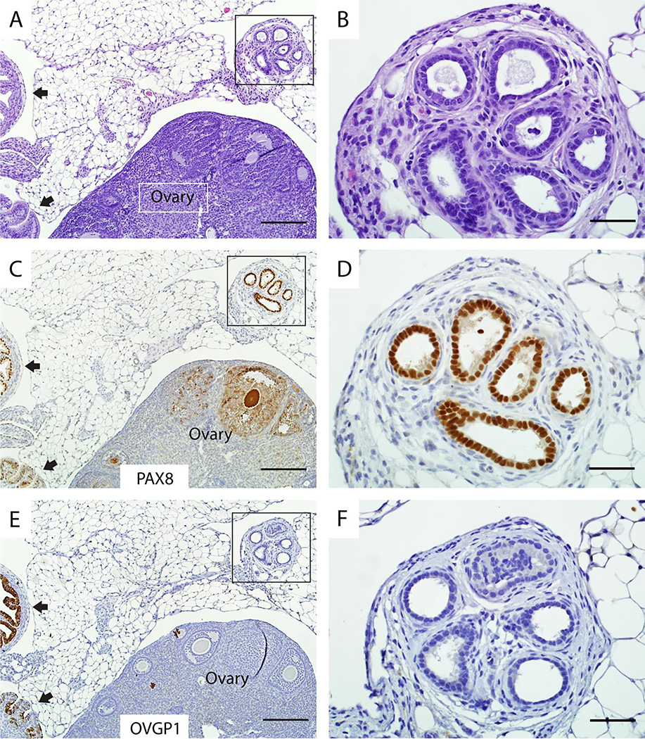
Figure 3. Endosalpingiosis lesions in Ovgp1-iCreERT2;R26RLSL-eYFP mice are different from rete ovarii.
Although extra-ovarian rete also forms glands lined by columnar cells (A) that express PAX8 (C), the rete does not express OVGP1 (E). High magnifications of boxed areas in A, C, and E are shown in B, D, F, respectively. Note the internal positive control for detection of PAX8 and OVGP1 expression (normal oviduct at black arrows) in lower left corners of panels C and E. Scale bars: 200 μm (panels A, C, E); 50 μm (panels B, D, F).

