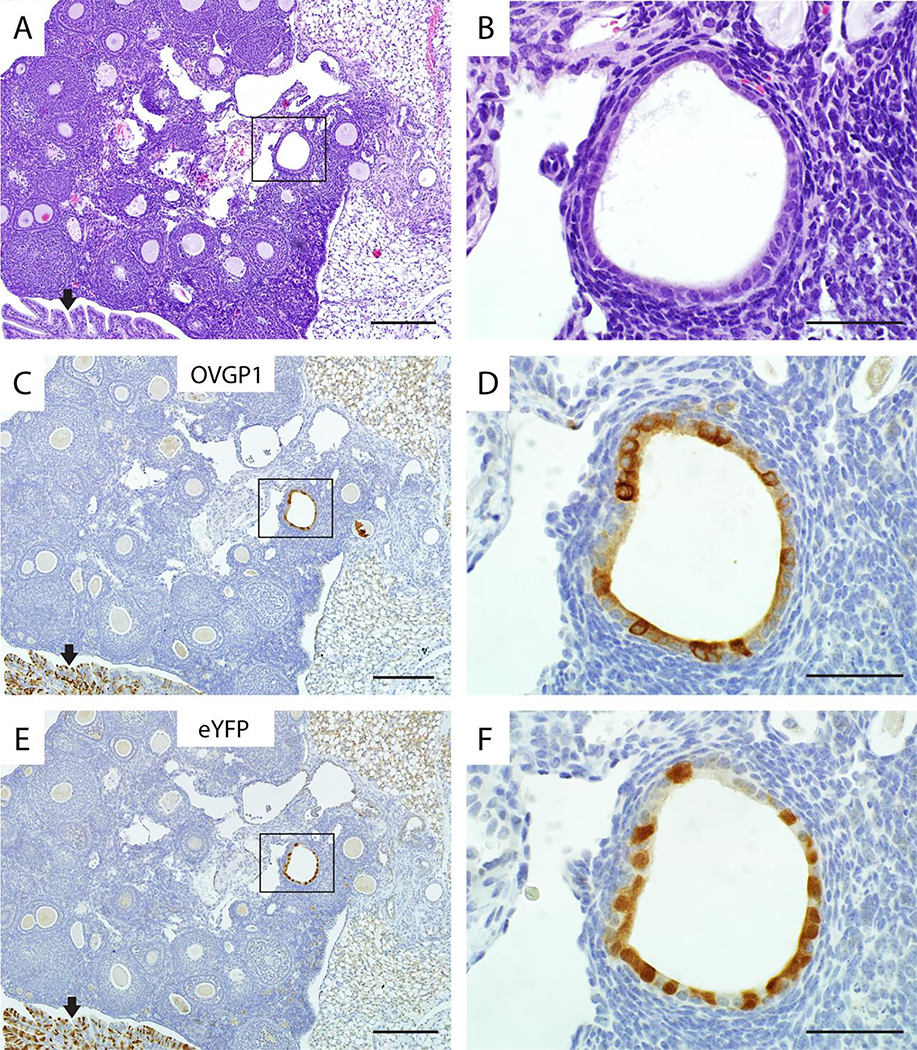
Figure 4. Ovarian endosalpingiosis lesions express both OVGP1 and eYFP in TAM-treated Ovgp1-iCreERT2;R26RLSL-eYFP mice.
A representative example of endosalpingiosis in a TAM-treated double transgenic mouse is shown: H&E stained section (A), and adjacent sections immunohistochemically stained for OVGP1 (C) and eYFP (E). High magnifications of boxed areas in panels A, C, and E are shown in panels B, D, and F, respectively. Note the internal positive control for detection of OVGP1 and eYFP expression (normal oviduct at black arrows) in lower left corners of panels C and E. Scale bars: 200 μm (panels A, C, E); 50 μm (panels B, D, F).

